BioMAX is the first X-ray macromolecular crystallography beamline of MAX IV Laboratory, which begins its user operations in 2017. It is a state-of-the-art resource accommodating multiple cutting edge experimental possibilities. The design goal for BioMAX was to create a stable and reliable beamline that is user friendly. The beamline experiment set-up is highly automated, in terms of both sample handling hardware and data analysis, including feedback on the data collection.
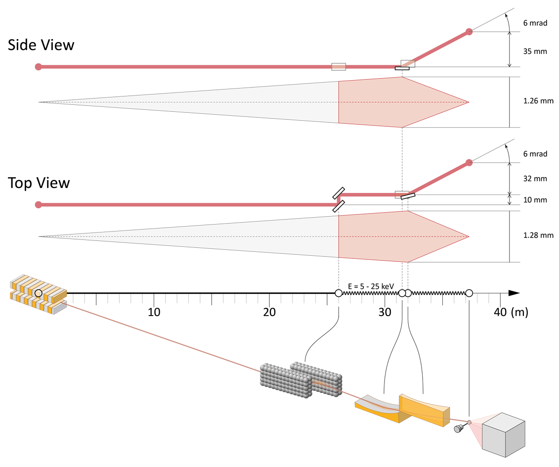
The X-ray beam focus is 20 x 5 μm2 at the sample position with a photon flux of 2 x 10¹³ ph/s at 500 mA ring current. The operational energy range of the beamline is 5–25 keV. Alternatively, using aperture overfilling it is possible to obtain a stable 5 x 5 μm2 beam at the sample position. Due to its extensive energy tunability, BioMAX is an ideal source for de novo phasing using the anomalous signal of heavy elements. Beam defocusing creates a practically parallel beam, which can be used to resolve extremely large unit cells > 1000 Å at high resolution. Due to its small beam cross-section and optional parallel beam, BioMAX is an optimal experimental set-up for X-ray crystallography using microcrystals and ultra large unit cells.
Endstation
Sample
The ISARA sample-changer dewar capacity: 29 UniPucks and 464 samples.
Techniques usage
Additional methods: MAD, SAD and SSAD.
Manipulator or Sample stage
Provides goniometry for the crystal samples.
Sample Environment
The ISARA sample changer provides cryogenic storage (LN2) of samples before measurements and transfer to the MD3 diffractometer.
The MD3 diffractometer can be equipped with a HC-Lab Humidity Controller.
Please contact staff for availability.
Dectris Eiger 16M
Max. frame rate 16M / 4M Mode: 133 Hz / 750 Hz
Detection
Fluorescence detector
Detection
- Crystallography (biological macromolecules)
- Molecular and cellular biology
Fotongatan 2
224 84 Lund
Sweden
- MXCuBE V3
- Diffraction data set
- HDF5