A) Beamline for spectroscopy of magnetic materials in the extreme ultraviolet spectral range
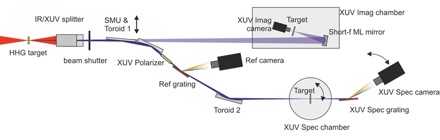
The extreme ultraviolet radiation (XUV) is generated in a standard gas (Ar, Ne, He) target. After a reflective XUV/IR beamsplitter the XUV light is focused by a toroidal mirror and circularly polarized by a 4-mirror reflective polarizer. We record a reference spectrum by diffracting the first order of a reflection grating by a in vacuum CCD camera. The 0 order is refocused by a second toroidal mirror onto the sampleto a spot size of approximately 100 µm FWHM. The transmitted or reflected light is collected by a flat field grating and collected by a second CCD camera. The two camera are synchronized and allow accurate and low noise measurements with an RMS ~ 1E-4. The sample environment includes an electromagnet to apply an external magnetic field of approximately 300 mT and a closed cycle, low vibration croystat for temperatures between ~10 K and 700 K.
Alternatively the polarization of the XUV beam can be controlled by a bi-circular pump field.
For a detailed overview please refer to a recent publication in Review of Scientific Instruments:
https://doi.org/10.1063/5.0013928
B) Beamline for coherent imaging and small angle scattering
By removing the switching mirror unit (SMU) the XUV beam is sent into a flexible vacuum chamber equipped with different monochromatizing focusing optics precise sample stages and a 4k in vacuum CCD camera.
Coherent Legend Elite Duo
For optical excitation there is currently the option to use the output from a optical parametric amplfier for accessing wavelength from 190 nm to 15 µm. Note that for extreme wavelength the pulse energy drops to approximately 1 µJ.
XUV spectrometer
Grazing incidence typ
lambda range: 11- 62 nm
sample - spectrometer: 350 mm
spectrometer - detector: 469 mm
https://www.hitachi-hightech.com/products/images/9797/ana-grating_05.pdf
Sample
Manipulator or Sample stage
Sample Environment
Magnetic Fields
CCD Camera
Type: Back-illuminated, enhanced process
AR Coating: No coating
Pixel Format: 1024 × 256
Image Area: 26.6mm × 6.7mm
Pixel Size: 26µm × 26µm
Full-Well Capacity: 500.000e -
Max. Dynamic Range: 125.000:1
Dark Current: 0.0005 eˉ/pixel/sec at -80°C
Camera features:
Flange: Vacuum flange ISO-F DN63
Read Noise at 500kHz: min 2.4eˉ rms, typical 4eˉ rms
Pixel Frequency: 250kHz – 3.0Mhz
ADC Resolution: 16 bit
CCD Sensor Cooling: -70°C to 20°C, forced air, water cooling
Interface: USB 2.0
Power Supply: 110-240VAC, max. 1A
Dimensions: 61mm × 89mm × 136mm
Detection
- Time-resolved studies
- XMCD
- Time-resolved studies
- Polarimetry
- Reflectrometry
- Time-resolved studies
- X-ray holography
- Coherent scattering
- Magnetic scattering
- Time-resolved scattering
- Knowledge based multifunctional materials
- Other - Material Sciences
- labview, spec
- dat
- h5, dat
- matlab, phython