Illuminating the molecular world with infrared brilliance
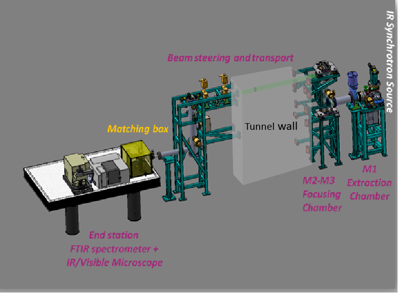
The SESAME BM02-IR beamline, operational since November 2018, is a cutting-edge facility serving the global Infrared scientific community with reliable capacity and advanced capabilities. The beamline was designed and built in collaboration with the French light source, SOLEIL. Dedicated for Synchrotron Radiation Fourier Transform Infrared Spectromicroscopy (SR-µFTIR), it uses a high-performance FTIR interferometer and harnesses infrared synchrotron radiation from both the bending magnet (BM) and edge radiation (ER) sources.
This vibrational technique, when coupled with synchrotron brilliance, delivers up to 1000× brighter signals than conventional thermal sources, with unique advantages in time structure, polarization, and broad wavelength coverage spanning the mid-IR (2.5–25 µm) and extending into the far-IR/THz region. The beamline provides powerful tools for non-destructive and spatially resolved chemical analysis. By integrating Fourier Transform Infrared (FTIR) spectrometry with advanced microscopy, the beamline enables scientists to identify and image IR-active vibrational modes at the diffraction-limited microscopic scale. In particular, it measures optically active molecular vibrations at the micro- and nano-scale, generating highly specific, spatially distributed chemical fingerprints. Diverse fields from material science and forensics, geology to mineralogy, food and agriculture, water and soil pollution and environmental studies, to pharma, biomedicine and life sciences, as well as cultural heritage, archaeology, palaeontology and art restoration.
With diffraction-limited spatial resolution and superior signal-to-noise ratios, the BM02-IR beamline opens new frontiers for discovery, offering scientists a powerful tool to explore the molecular world in unprecedented detail.
SESAME Joins INFN-CHNet with New Cultural Heritage Laboratory
In 2022, SESAME inaugurated a new laboratory dedicated to cultural heritage as part of the Cultural Heritage Network, CHNet, of the Italian National Institute for Nuclear Physics (INFN). This important milestone marked SESAME’s official inclusion in the network, reinforcing international collaboration. To support advanced research, INFN has equipped the BM02-IR beamline with a state-of-the-art endstation providing powerful tools for non-invasive analysis, enabling researchers to explore and safeguard cultural treasures.
BM D02
Offline-mode Endstation (Globar IR Source)
The microscope features Kohler Illumination for Reflection and Transmission, Brightfield, Darkfield and Polarized Light. In addition to DIC (Differential Interference Contrast) Optics; and Fluorescence illumination of:
-Wide Band Blue Fluorescence Cube 450-480nm,
-Wide Band Green Fluorescence Cube 510-550nm,
-Wide Band UV Fluorescence Cube 330-385nm.
DTGS
InGaAs
Sample
Specac Mini Pellet Press™ | FTIR transmission mode,
Specac Micro Compression Cell™ | FTIR transmission mode,
Specac DC-3 | Diamond Compression Cell™ | FTIR microscopy in transmission mode,
Specac Monolayer Grazing Angle Specular Reflectance Accessory™ | FTIR specular reflection mode,
Pike Technologies MIRacle™ cell | FTIR single reflection mode,
Thermo Fisher Slide-on 15x ATR Si Objective™
Manipulator or Sample stage
Sample Holders
Specac Mini Pellet Press™ | FTIR transmission mode,
Specac Micro Compression Cell™ | FTIR transmission mode,
Specac DC-3 | Diamond Compression Cell™ | FTIR microscopy in transmission mode,
Specac Monolayer Grazing Angle Specular Reflectance Accessory™ | FTIR specular reflection mode,
Pike Technologies MIRacle™ cell | FTIR single reflection mode,
Thermo Slide-on 15x ATR Si Objective™
Standard SR-IR Endstation
DTGS
InGaAs
Focal Plane Array (FPA)
Sample
Specac Mini Pellet Press™ | FTIR transmission mode,
Specac Micro Compression Cell™ | FTIR transmission mode,
Specac DC-3 | Diamond Compression Cell™ | FTIR microscopy in transmission mode,
Specac Monolayer Grazing Angle Specular Reflectance Accessory™ | FTIR specular reflection mode,
Pike Technologies MIRacle™ cell | FTIR single reflection mode,
Bruker 20x ATR Ge Objective™
Sample Holders
Specac Mini Pellet Press™ | FTIR transmission mode,
Specac Micro Compression Cell™ | FTIR transmission mode,
Specac DC-3 | Diamond Compression Cell™ | FTIR microscopy in transmission mode,
Specac Monolayer Grazing Angle Specular Reflectance Accessory™ | FTIR specular reflection mode,
Pike Technologies MIRacle™ cell | FTIR single reflection mode,
Bruker 20x ATR Ge Objective™
DTGS
- TE Cooled DLaTGS Detector with KBr Window (12,500-350 cm-1);
- DLaTGS Detector with Polyethylene Window (700-50 cm-1).
Detection
Focal Plane Array (FPA)
Detection
InGaAs
InGaAs detector for NIR (12.000-3.800 cm-1).
Detection
MCT
- 50um MCT-A Detector;
- MCT-B Detector.
Detection
- IR spectroscopy
- IR Microscopy
- Biochemistry
- Catalysis
- Electrochemistry
- Green Chemistry
- Technique Development - Chemistry
- Geology
- Marine science/Oceanography
- Mineralogy
- Other - Earth Sciences & Environment
- Plant science
- Technique Development - Earth Sciences & Environment
- Water sciences/Hydrology
- Sustainable energy systems
- Technique Development - Energy
- Detectors
- Nanotechnology & production processes
- Other - Engineering & Technology
- Space
- Technique Development - Engineering & Technology
- Arts
- Cultural Heritage
- Components & Micro-systems
- Biophysics
- Food quality and safety
- Medicine
- Molecular and cellular biology
- Other - Life Sciences & Biotech
- Pharmaceutics (drug screening)
- Technique Development - Life Sciences & Biotech
- Veterinary sciences
- Knowledge based multifunctional materials
- Metallurgy
- Other - Material Sciences
- Technique Development - Material Sciences
- Astronomy/Astrophysics/Astroparticles
- Atomic & molecular physics
- Optics
- Soft condensed matter physics
- Surfaces, interfaces and thin films
- Technique Development - Physics
P.O Box (7) Allan (19252) Jordan
- Beamline Control: EPICS (EPICS - Experimental Physics and Industrial Control System)
Beamline Standard Endstation: OPUS® 8.5 (SP1) Build 8,7,10 Proprietary software (Copyright © Bruker Optik GmbH 2020).
- Transmittance/Reflectance/Absorbance IR single spectra, interferograms, visible images, and hyperspectral images/chemical maps.
- Standard Endstation: OPUS files as single spectra and maps (possible conversion to other formats.),
Offline Endstation: OMNIC as single spectra and maps (possible conversion to other formats.)
- On-site and remote access to:
OPUS® 8.5 (SP1) Build 8,7,10 (Copyright © Bruker Optik GmbH 2020),
OMNIC® 9.2.41, OMNICMC® v 9.1.0, OMNIC Atlus® 9.1.24. (Copyright © 1992-2012 Thermo Fisher Scientific Inc.),
The Unscrambler X® v 10.4. (Copyright © CAMO Analytics),
CytoSpec® v 1.3.02. (Copyright © 2000-2020 Peter Lasch),
PeakFit v4.12 (Systat Software, USA),
Quasar®.